
The open fruit fly brain map: what Google and Janelia released
Google Research and HHMI Janelia published a complete connectome of the adult male fruit fly central nervous system. Learn what the open dataset contains, what it does not, and why it matters for AI-assisted science.

What Google and Janelia released
On September 3, 2026, Google Research announced the publication of a complete wiring map of the adult male fruit fly's brain and central nervous system. The project was led by HHMI Janelia with Google Research, the Cambridge Drosophila Connectomics Group, and other collaborators.
The scientific term for this map is a connectome. A connectome records neurons and the connections between them. In this case, it covers the central brain, optic lobes, neck connective, and ventral nerve cord, which performs some functions comparable to a spinal cord.
Google Research's announcement describes more than 166,000 neurons and 125 million synaptic connections. The final Cell paper reports 166,700 reconstructed neurons and 124.2 million contacts between proofread neurons. The difference reflects the level of precision and the counting definition used by each source.
The dataset timeline also matters. The MaleCNS release notes record version 1.0 on June 8, 2026. The paper and Google's public announcement followed on September 3.
It is an open research system, not one application
Some online descriptions call the release a Google GitHub repository. That is convenient shorthand, but it misses how the project is organized.
The public resource is the Male CNS connectome dataset, hosted through HHMI Janelia. Researchers can download image volumes, neuron annotations, connectivity tables, and related data. The dataset is available under a Creative Commons Attribution 4.0 licence.
The paper-specific GitHub repository contains supporting analysis and supplemental material. It is not a single application that installs the complete research project.
Researchers can explore the data through several interfaces. These include neuPrint for connectivity queries, the MaleCNS Cell Type Explorer, programmatic Python and R access, and Neuroglancer.
Neuroglancer is Google's open-source, browser-based viewer for large volumetric datasets. It can display cross-sections, three-dimensional meshes, and neuron skeletons. The viewer predates the MaleCNS release and is part of the infrastructure that makes the dataset usable. It is not a brain simulator.
AI helped turn tissue images into a usable map
Producing a connectome begins with extremely thin sections of biological tissue. Electron microscopes capture images of those sections. Researchers then identify cells and reconstruct their shapes across a large image volume.
Google's connectomics researchers use AI systems to help turn two-dimensional microscope images into three-dimensional neural structures. Techniques such as flood-filling networks identify pixels that appear to belong to the same biological object and connect them across the image volume.
AI did not remove the need for scientific review. Human experts at Janelia proofread and annotated the male fruit fly connectome before publication.
This combination is one of the most useful lessons from the project. Automation handled image-processing work at a scale that would be difficult to approach manually. Domain experts remained responsible for verification, annotation, and scientific meaning.
What the map does not represent
A connectome is not a digital consciousness or a complete simulation of an animal.
It describes physical connections between neurons. It does not, by itself, reproduce every electrical signal, chemical process, biological state, memory, or environmental influence involved in behaviour.
Developers may connect parts of the dataset to simulations, games, or experimental control systems. Those demonstrations are downstream interpretations. They should not be treated as evidence that a living brain has been uploaded or recreated in software.
Clear language matters when scientific work becomes a viral technology story. "Scientists published an open neural wiring map" is accurate. "Google uploaded a fly's mind" is not.
Why the open release matters
The immediate value is scientific. Researchers can study how sensory inputs connect to movement, compare male and female nervous systems, investigate differences between individuals, and test hypotheses about vision, taste, courtship, aggression, and other behaviours.
The wider lesson concerns research infrastructure. Useful open systems provide more than a downloadable file. They connect several layers:
- A documented source dataset.
- A stable representation of the data.
- Tools for exploration and programmatic access.
- Human-reviewed annotations.
- Versioning, licensing, and publication records.
- A community that can reproduce, challenge, and extend the work.
This structure allows independent teams to ask new questions without recreating the entire data pipeline.
A practical lesson for AI teams
Organizations adopting AI can apply the same discipline on a smaller scale. Before calling a system open, intelligent, or production-ready, ask:
- What has actually been released: code, model weights, data, an interface, or a combination of them?
- What does the artifact represent, and what remains outside it?
- Which parts were generated automatically?
- Which parts were reviewed by qualified people?
- What licence and version govern reuse?
- What evidence would another team need to reproduce the result?
- Which claims are demonstrated, and which remain experimental?
These questions replace spectacle with evidence. They also help teams distinguish a compelling demonstration from a dependable operating resource.
The real breakthrough is the shared foundation
The male fruit fly connectome is remarkable because of its scale, but its openness may prove just as important.
Researchers now have a common, inspectable foundation for studying how a nervous system connects perception to action. The dataset, interfaces, open-source viewing tools, and human verification make further work possible.
That is the deeper lesson for AI-assisted science. Progress rarely comes from a model acting alone. It comes from building reliable systems in which data, software, expertise, and verification reinforce one another.
Image and data credit
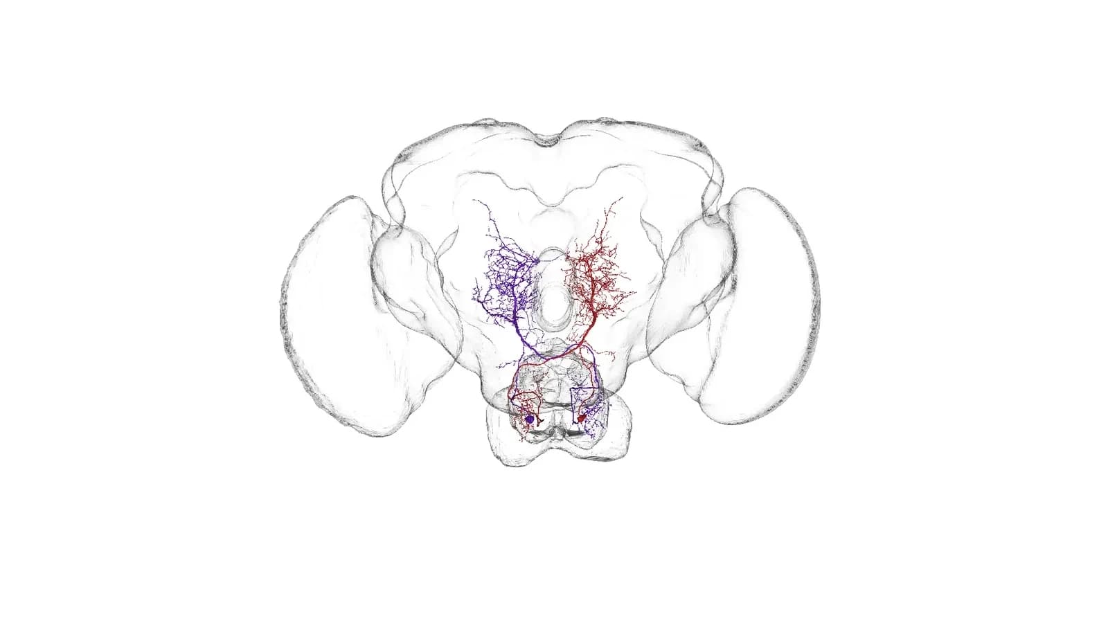
The hero shows two DNg13 descending-neuron reconstructions from the MaleCNS dataset. Data credit: FlyEM at HHMI Janelia, University of Cambridge, MRC Laboratory of Molecular Biology, Google Research, and the contributors named by Berg and collaborators. The data is licensed under CC BY 4.0. The visualization was rendered in Google Neuroglancer and sourced from Harshith Vaddiparthy's documented exploration. Kenzai added neutral canvas space and created a social crop without changing the neuron geometry.